Brain Anatomy Ct Scan Images . when reading a brain computed tomography (ct), the left side of the image depicts the patient’s right side,. Ct brain by gourab mitro plaban. The cerebral cortex is a layer of grey matter formed in gyri (folds) over the entire brain surface. Citation, doi, disclosures and article data. Annotated teaching ct head in standard and bone windows. important grey matter structures visible on ct images of the brain include the cortex, insula, basal ganglia, and thalamus. This article lists a series of labeled imaging anatomy. this tutorial takes you through the important anatomy required to understand ct images of the brain. Hover on/off image to show/hide findings. Ct head by mohit kumar. 111 normal anatomy by mohamed shweel. 78 public playlists include this case.
from flickr.com
Ct head by mohit kumar. This article lists a series of labeled imaging anatomy. when reading a brain computed tomography (ct), the left side of the image depicts the patient’s right side,. Ct brain by gourab mitro plaban. Citation, doi, disclosures and article data. 78 public playlists include this case. 111 normal anatomy by mohamed shweel. this tutorial takes you through the important anatomy required to understand ct images of the brain. important grey matter structures visible on ct images of the brain include the cortex, insula, basal ganglia, and thalamus. Hover on/off image to show/hide findings.
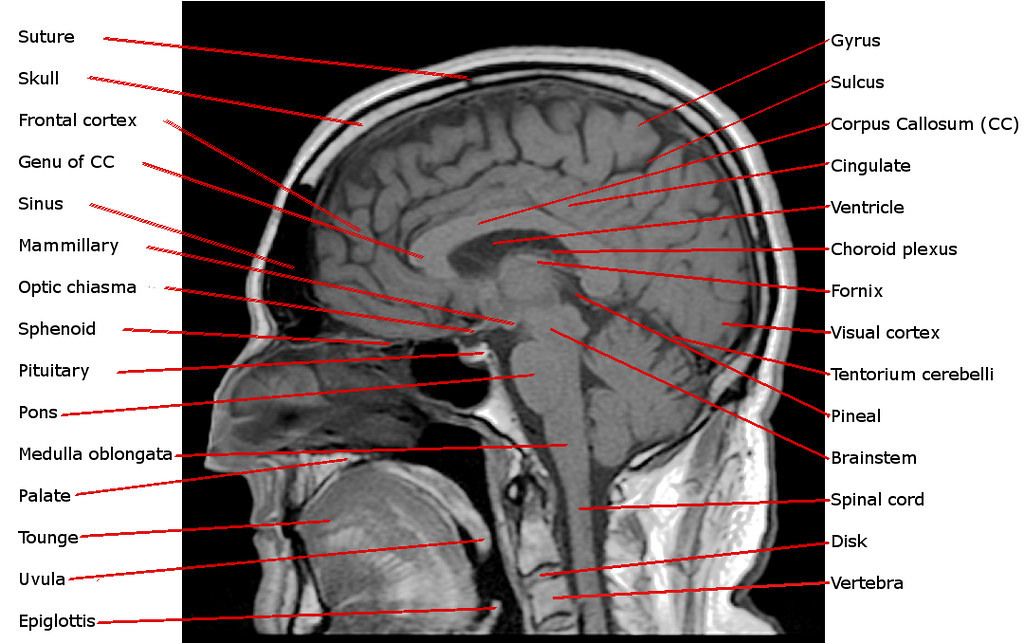
Annotated Sagittal T1 Midline MRI Scan of Reigh's Brain Flickr
Brain Anatomy Ct Scan Images when reading a brain computed tomography (ct), the left side of the image depicts the patient’s right side,. this tutorial takes you through the important anatomy required to understand ct images of the brain. 78 public playlists include this case. Hover on/off image to show/hide findings. Citation, doi, disclosures and article data. Ct brain by gourab mitro plaban. Ct head by mohit kumar. when reading a brain computed tomography (ct), the left side of the image depicts the patient’s right side,. important grey matter structures visible on ct images of the brain include the cortex, insula, basal ganglia, and thalamus. The cerebral cortex is a layer of grey matter formed in gyri (folds) over the entire brain surface. Annotated teaching ct head in standard and bone windows. This article lists a series of labeled imaging anatomy. 111 normal anatomy by mohamed shweel.
From
Brain Anatomy Ct Scan Images when reading a brain computed tomography (ct), the left side of the image depicts the patient’s right side,. The cerebral cortex is a layer of grey matter formed in gyri (folds) over the entire brain surface. This article lists a series of labeled imaging anatomy. Ct brain by gourab mitro plaban. Ct head by mohit kumar. Citation, doi, disclosures. Brain Anatomy Ct Scan Images.
From
Brain Anatomy Ct Scan Images The cerebral cortex is a layer of grey matter formed in gyri (folds) over the entire brain surface. 78 public playlists include this case. This article lists a series of labeled imaging anatomy. important grey matter structures visible on ct images of the brain include the cortex, insula, basal ganglia, and thalamus. when reading a brain computed tomography. Brain Anatomy Ct Scan Images.
From
Brain Anatomy Ct Scan Images this tutorial takes you through the important anatomy required to understand ct images of the brain. Annotated teaching ct head in standard and bone windows. important grey matter structures visible on ct images of the brain include the cortex, insula, basal ganglia, and thalamus. when reading a brain computed tomography (ct), the left side of the image. Brain Anatomy Ct Scan Images.
From
Brain Anatomy Ct Scan Images this tutorial takes you through the important anatomy required to understand ct images of the brain. Citation, doi, disclosures and article data. important grey matter structures visible on ct images of the brain include the cortex, insula, basal ganglia, and thalamus. This article lists a series of labeled imaging anatomy. Ct brain by gourab mitro plaban. The cerebral. Brain Anatomy Ct Scan Images.
From
Brain Anatomy Ct Scan Images important grey matter structures visible on ct images of the brain include the cortex, insula, basal ganglia, and thalamus. 111 normal anatomy by mohamed shweel. this tutorial takes you through the important anatomy required to understand ct images of the brain. Annotated teaching ct head in standard and bone windows. Hover on/off image to show/hide findings. This. Brain Anatomy Ct Scan Images.
From
Brain Anatomy Ct Scan Images 78 public playlists include this case. this tutorial takes you through the important anatomy required to understand ct images of the brain. Annotated teaching ct head in standard and bone windows. when reading a brain computed tomography (ct), the left side of the image depicts the patient’s right side,. Ct head by mohit kumar. important grey matter. Brain Anatomy Ct Scan Images.
From radiology.ucsf.edu
Exploring the Brain How Are Brain Images Made with CT? UCSF Radiology Brain Anatomy Ct Scan Images Ct head by mohit kumar. Citation, doi, disclosures and article data. this tutorial takes you through the important anatomy required to understand ct images of the brain. important grey matter structures visible on ct images of the brain include the cortex, insula, basal ganglia, and thalamus. 78 public playlists include this case. This article lists a series of. Brain Anatomy Ct Scan Images.
From
Brain Anatomy Ct Scan Images The cerebral cortex is a layer of grey matter formed in gyri (folds) over the entire brain surface. Annotated teaching ct head in standard and bone windows. 78 public playlists include this case. This article lists a series of labeled imaging anatomy. 111 normal anatomy by mohamed shweel. Ct brain by gourab mitro plaban. Citation, doi, disclosures and article. Brain Anatomy Ct Scan Images.
From
Brain Anatomy Ct Scan Images 78 public playlists include this case. Ct brain by gourab mitro plaban. important grey matter structures visible on ct images of the brain include the cortex, insula, basal ganglia, and thalamus. Ct head by mohit kumar. Annotated teaching ct head in standard and bone windows. 111 normal anatomy by mohamed shweel. when reading a brain computed tomography. Brain Anatomy Ct Scan Images.
From
Brain Anatomy Ct Scan Images 78 public playlists include this case. Ct head by mohit kumar. The cerebral cortex is a layer of grey matter formed in gyri (folds) over the entire brain surface. this tutorial takes you through the important anatomy required to understand ct images of the brain. Citation, doi, disclosures and article data. 111 normal anatomy by mohamed shweel. . Brain Anatomy Ct Scan Images.
From
Brain Anatomy Ct Scan Images 111 normal anatomy by mohamed shweel. Annotated teaching ct head in standard and bone windows. Citation, doi, disclosures and article data. this tutorial takes you through the important anatomy required to understand ct images of the brain. Ct brain by gourab mitro plaban. important grey matter structures visible on ct images of the brain include the cortex,. Brain Anatomy Ct Scan Images.
From
Brain Anatomy Ct Scan Images Ct brain by gourab mitro plaban. Annotated teaching ct head in standard and bone windows. 111 normal anatomy by mohamed shweel. this tutorial takes you through the important anatomy required to understand ct images of the brain. important grey matter structures visible on ct images of the brain include the cortex, insula, basal ganglia, and thalamus. Ct. Brain Anatomy Ct Scan Images.
From www.casestacks.com
MRI Brain Anatomy Brain Anatomy Ct Scan Images 78 public playlists include this case. Ct head by mohit kumar. this tutorial takes you through the important anatomy required to understand ct images of the brain. This article lists a series of labeled imaging anatomy. important grey matter structures visible on ct images of the brain include the cortex, insula, basal ganglia, and thalamus. Annotated teaching ct. Brain Anatomy Ct Scan Images.
From learningneurology.com
Approach to CT head Brain Anatomy Ct Scan Images 111 normal anatomy by mohamed shweel. when reading a brain computed tomography (ct), the left side of the image depicts the patient’s right side,. 78 public playlists include this case. Ct brain by gourab mitro plaban. Hover on/off image to show/hide findings. This article lists a series of labeled imaging anatomy. Ct head by mohit kumar. Annotated teaching. Brain Anatomy Ct Scan Images.
From
Brain Anatomy Ct Scan Images 78 public playlists include this case. Citation, doi, disclosures and article data. The cerebral cortex is a layer of grey matter formed in gyri (folds) over the entire brain surface. 111 normal anatomy by mohamed shweel. Ct head by mohit kumar. Ct brain by gourab mitro plaban. important grey matter structures visible on ct images of the brain. Brain Anatomy Ct Scan Images.
From pn.bmj.com
Normal anatomy of the brain on CT and MRI with a few normal variants Brain Anatomy Ct Scan Images Hover on/off image to show/hide findings. Annotated teaching ct head in standard and bone windows. 111 normal anatomy by mohamed shweel. This article lists a series of labeled imaging anatomy. 78 public playlists include this case. The cerebral cortex is a layer of grey matter formed in gyri (folds) over the entire brain surface. this tutorial takes you. Brain Anatomy Ct Scan Images.
From www.bjaed.org
Brain imaging for anaesthetists and intensivists part Brain Anatomy Ct Scan Images when reading a brain computed tomography (ct), the left side of the image depicts the patient’s right side,. important grey matter structures visible on ct images of the brain include the cortex, insula, basal ganglia, and thalamus. The cerebral cortex is a layer of grey matter formed in gyri (folds) over the entire brain surface. Hover on/off image. Brain Anatomy Ct Scan Images.
From www.southsudanmedicaljournal.com
How to interpret an unenhanced CT Brain scan. Part 1 Basic principles Brain Anatomy Ct Scan Images Citation, doi, disclosures and article data. 111 normal anatomy by mohamed shweel. 78 public playlists include this case. This article lists a series of labeled imaging anatomy. Ct head by mohit kumar. important grey matter structures visible on ct images of the brain include the cortex, insula, basal ganglia, and thalamus. Ct brain by gourab mitro plaban. Hover. Brain Anatomy Ct Scan Images.